 Movie
Movie Controller
 Structure viewers
 About EMN search


-Search query
-Search result
Showing 1 - 50 of 89 items for (author: atherton & j)

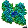
EMDB-42681: 
The structure of the native cardiac thin filament troponin core in Ca2+-free state from the upper strand
Method: single particle / : Galkin VE, Risi CM

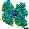
EMDB-42682:
The structure of the native cardiac thin filament troponin core in Ca2+-free tilted state from the upper strand
Method: single particle / : Galkin VE, Risi CM
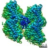
EMDB-42683:
The structure of the native cardiac thin filament troponin core in Ca2+-free rotated state from the upper strand
Method: single particle / : Galkin VE, Risi CM

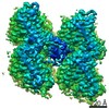
EMDB-42800:
The structure of the native cardiac thin filament troponin core in Ca2+-free state from the lower strand
Method: single particle / : Galkin VE, Risi CM

EMDB-42833:
The structure of the native cardiac thin filament troponin core in Ca2+-free rotated state from the lower strand
Method: single particle / : Galkin VE, Risi CM

EMDB-42835:
The structure of the native cardiac thin filament troponin core in Ca2+-free tilted state from the lower strand
Method: single particle / : Galkin VE, Risi CM

EMDB-42846:
The structure of the native cardiac thin filament troponin core in Ca2+-bound fully activated state from the upper strand
Method: single particle / : Galkin VE, Risi CM

EMDB-42847:
The structure of the native cardiac thin filament troponin core in Ca2+-bound partially activated state from the upper strand
Method: single particle / : Galkin VE, Risi CM

EMDB-42849:
The structure of the native cardiac thin filament troponin core in Ca2+-bound fully activated state 1 from the lower strand
Method: single particle / : Galkin VE, Risi CM

EMDB-42856:
The structure of the native cardiac thin filament troponin core in Ca2+-bound fully activated state 2 from the lower strand
Method: single particle / : Galkin VE, Risi CM

EMDB-42858:
The structure of the native cardiac thin filament troponin core in Ca2+-bound partially activated state from the lower strand
Method: single particle / : Galkin VE, Risi CM

EMDB-42874:
The structure of the native cardiac thin filament troponin core in Ca2+-free state from the upper strand activated by the C1-domain of cardiac myosin binding protein C
Method: single particle / : Galkin VE, Risi CM

PDB-8uww:
The structure of the native cardiac thin filament troponin core in Ca2+-free state from the upper strand
Method: single particle / : Galkin VE, Risi CM

PDB-8uwx:
The structure of the native cardiac thin filament troponin core in Ca2+-free tilted state from the upper strand
Method: single particle / : Galkin VE, Risi CM
PDB-8uwy:
The structure of the native cardiac thin filament troponin core in Ca2+-free rotated state from the upper strand
Method: single particle / : Galkin VE, Risi CM

PDB-8uyd:
The structure of the native cardiac thin filament troponin core in Ca2+-free state from the lower strand
Method: single particle / : Galkin VE, Risi CM

PDB-8uz5:
The structure of the native cardiac thin filament troponin core in Ca2+-free rotated state from the lower strand
Method: single particle / : Galkin VE, Risi CM

PDB-8uz6:
The structure of the native cardiac thin filament troponin core in Ca2+-free tilted state from the lower strand
Method: single particle / : Galkin VE, Risi CM

PDB-8uzx:
The structure of the native cardiac thin filament troponin core in Ca2+-bound fully activated state from the upper strand
Method: single particle / : Galkin VE, Risi CM

PDB-8uzy:
The structure of the native cardiac thin filament troponin core in Ca2+-bound partially activated state from the upper strand
Method: single particle / : Galkin VE, Risi CM

PDB-8v01:
The structure of the native cardiac thin filament troponin core in Ca2+-bound fully activated state 1 from the lower strand
Method: single particle / : Galkin VE, Risi CM

PDB-8v0i:
The structure of the native cardiac thin filament troponin core in Ca2+-bound fully activated state 2 from the lower strand
Method: single particle / : Galkin VE, Risi CM

PDB-8v0k:
The structure of the native cardiac thin filament troponin core in Ca2+-bound partially activated state from the lower strand
Method: single particle / : Galkin VE, Risi CM

PDB-8v0y:
The structure of the native cardiac thin filament troponin core in Ca2+-free state from the upper strand activated by the C1-domain of cardiac myosin binding protein C
Method: single particle / : Galkin VE, Risi CM

EMDB-14386:
In situ subtomogram average of long-repeat F-actin from neuronal growth cones
Method: subtomogram averaging / : Atherton J, Stouffer M, Francis F, Moores CA

EMDB-14395:
In situ subtomogram average of short-repeat F-actin from neuronal growth cones
Method: subtomogram averaging / : Atherton J, Stouffer M, Francis F, Moores CA

EMDB-14396:
Tomogram of wild-type mouse neuronal growth cone (T-zone), 4 x binned, processed.
Method: electron tomography / : Atherton J, Stouffer M, Francis F, Moores CA

EMDB-14397:
Tomogram of wild-type mouse neuronal growth cone (P-zone), 4 x binned, processed.
Method: electron tomography / : Atherton J, Stouffer M, Francis F, Moores CA

EMDB-14400:
Tomogram of wild-type mouse neuronal growth cone (C-zone), 4 x binned, processed.
Method: electron tomography / : Atherton J, Stouffer M, Francis F, Moores CA

EMDB-14401:
Tomogram of doublecortin knock-out mouse neuronal growth cone (C-zone), 4 x binned, processed.
Method: electron tomography / : Atherton J, Stouffer M, Francis F, Moores CA

EMDB-14402:
Tomogram of doublecortin knock-out mouse neuronal growth cone (P-zone), 4 x binned, processed.
Method: electron tomography / : Atherton J, Stouffer M, Francis F, Moores CA

EMDB-14414:
Tomogram of doublecortin knock-out mouse neuronal growth cone (T-zone), 4 x binned, processed.
Method: electron tomography / : Atherton J, Stouffer M, Francis F, Moores CA

EMDB-14416:
Tomogram of wild-type mouse neuronal growth cone (P-zone), 2 x binned, processed.
Method: electron tomography / : Atherton J, Stouffer M, Francis F, Moores CA

EMDB-12257:
Plasmodium falciparum kinesin-5 motor domain without nucleotide, complexed with 14 protofilament microtubule.
Method: helical / : Cook AD, Roberts A, Atherton J, Tewari R, Topf M, Moores CA

EMDB-12258:
Plasmodium falciparum kinesin-5 motor domain bound to AMPPNP, complexed with 14 protofilament microtubule.
Method: helical / : Cook AD, Roberts A, Atherton J, Tewari R, Topf M, Moores CA

PDB-7nb8:
Plasmodium falciparum kinesin-5 motor domain without nucleotide, complexed with 14 protofilament microtubule.
Method: helical / : Cook AD, Roberts A, Atherton J, Tewari R, Topf M, Moores CA

PDB-7nba:
Plasmodium falciparum kinesin-5 motor domain bound to AMPPNP, complexed with 14 protofilament microtubule.
Method: helical / : Cook AD, Roberts A, Atherton J, Tewari R, Topf M, Moores CA

EMDB-11338:
Kinesin binding protein (KBP)
Method: single particle / : Atherton J, Hummel JJA

EMDB-11339:
Kinesin binding protein complexed with Kif15 motor domain
Method: single particle / : Atherton J, Hummel JJA
EMDB-11340:
Microtubule complexed with Kif15 motor domain. Symmetrised asymmetric unit
Method: single particle / : Atherton J, Hummel JJA, Olieric N, Locke J, Pena A, Rosenfeld SS, Steinmetz MO, Hoogenraad CC, Moores CA

PDB-6zpg:
Kinesin binding protein (KBP)
Method: single particle / : Atherton J, Hummel JJA, Olieric N, Locke J, Pena A, Rosenfeld SS, Steinmetz MO, Hoogenraad CC, Moores CA
PDB-6zph:
Kinesin binding protein complexed with Kif15 motor domain
Method: single particle / : Atherton J, Hummel JJA, Olieric N, Locke J, Pena A, Rosenfeld SS, Steinmetz MO, Hoogenraad CC, Moores CA

PDB-6zpi:
Microtubule complexed with Kif15 motor domain. Symmetrised asymmetric unit
Method: single particle / : Atherton J, Hummel JJA, Olieric N, Locke J, Pena A, Rosenfeld SS, Steinmetz MO, Hoogenraad CC, Moores CA
EMDB-4862:
Cryo-EM structure of the N-terminal DC repeat (NDC) of NDC-NDC chimera (human sequence) bound to 13-protofilament GDP-microtubule
Method: single particle / : Manka SW

PDB-6rf8:
Cryo-EM structure of the N-terminal DC repeat (NDC) of NDC-NDC chimera (human sequence) bound to 13-protofilament GDP-microtubule
Method: single particle / : Manka SW
EMDB-4643:
HsCKK (human CAMSAP1) decorated 13pf taxol-GDP microtubule
Method: single particle / : Atherton JM, Luo Y, Xiang S, Yang C, Jiang K, Stangier M, Vemu A, Cook A, Wang S, Roll-Mecak A, Steinmetz MO, Akhmanova A, Baldus M, Moores CA
EMDB-4644:
NgCKK (N.Gruberi CKK) decorated 13pf taxol-GDP microtubule
Method: single particle / : Atherton JM, Luo Y, Xiang S, Yang C, Jiang K, Stangier M, Vemu A, Cook A, Wang S, Roll-Mecak A, Steinmetz MO, Akhmanova A, Baldus M, Moores CA
EMDB-4650:
NgCKK (Naegleria Gruberi CKK) decorated 14pf taxol-GDP microtubule
Method: single particle / : Atherton JM, Luo Y, Xiang S, Yang C, Jiang K, Stangier M, Vemu A, Cook A, Wang S, Roll-Mecak A, Steinmetz MO, Akhmanova A, Baldus M, Moores CA
EMDB-4654:
HsCKK (human CAMSAP1) decorated 14pf taxol-GDP microtubule
Method: single particle / : Atherton JM, Luo Y, Xiang S, Yang C, Jiang K, Stangier M, Vemu A, Cook A, Wang S, Roll-Mecak A, Steinmetz MO, Akhmanova A, Baldus M, Moores CA
PDB-6qus:
HsCKK (human CAMSAP1) decorated 13pf taxol-GDP microtubule
Method: single particle / : Atherton JM, Luo Y, Xiang S, Yang C, Jiang K, Stangier M, Vemu A, Cook A, Wang S, Roll-Mecak A, Steinmetz MO, Akhmanova A, Baldus M, Moores CA
Pages:
 wwPDB to switch to version 3 of the EMDB data model
